


Effects of High Hydrostatic Pressure on Proteins
-
摘要: 在现有的超高压对蛋白质影响研究的基础上,详细地总结了超高压对蛋白质的分子体积、非共价键和分子结构的影响。在超高压作用下,蛋白质的分子体积被压缩变小;压力通过改变蛋白质分子的氢键、离子键、水合作用和疏水相互作用来影响蛋白质结构;低于800 MPa的压力会造成蛋白质分子的二级、三级和四级结构的改变,其中四级结构对压力最敏感,三级结构次之,二级结构的改变较小;高于8 GPa的压力会影响蛋白质分子的一级结构。Abstract: This review introduces the effects of high hydrostatic pressure on the molecular volume, non-covalent bond and conformation of protein.The molecular volume is decreased, and the hydrogen bond, electrovalent bond, and hydrophobic interaction are influenced by compression.The secondary, tertiary and quaternary structures of protein are influenced by pressure below 800 MPa.Tertiary and quaternary structures are more pressure-sensitive than secondary structure.Pressure below 8 GPa does not influence the primary structure of proteins.
-
Key words:
- ultra-high pressure /
- protein /
- molecular volume /
- non-covalent bond /
- conformation
-
表 1 超高压对蛋白质分子体积的影响
Table 1. Influences of ultra-high pressure on the molecular volume of proteins
Protein Pressure/(MPa) Molecular volume changes Test method Apomyoglobin 250 -61 mL/mol[15] Fluorescence Staphylococcal nuclease 100 -50 mL/mol[16] DSC Staphylococcal nuclease 300 -69--104 mL/mol[17] NMR, Fluorescence Bovine pancreatic ribonuclease 250 -21 mL/mol[14] NMR Flavodoxin from Peptostreptococcus elsdenii 870 -74 mL/mol[18] Fluorescence Flavodoxin from Desulfovibrio vulgaris 470 -63 mL/mol[18] Fluorescence Flavodoxin from Azotobacter vinelandii 1 060 -64 mL/mol[18] Fluorescence Metmyoglobin 60-600 -51--114 mL/mol[19] DSC, UV spectrum Cytochrome oxidase 250 -80 mL/mol[20] DSC, Visible spectrum Cytochrome b562 110 -102 mL/mol[21] NMR T4 lysozyme 200 -0.194 nm3[22] NMR, High pressure crystallography T4 lysozyme 50 -0.210 nm3[22] NMR, High pressure crystallography Note:DSC means differential scanning calorimetry; NMR means nuclear magnetic resonance.  下载: 导出CSV
表 2 超高压对蛋白质二级结构的影响
Table 2. Influences of ultra-high pressure on the secondary structure of proteins
Protein Pressure/(MPa) Changes of secondary structure Test method β-lactoglobulin 140 No change[43] FTIR β-lactoglobulin 600 The contents of α-helix and β-sheet decrease from
41% and 34% to 34% and 30%, respectively; The
content of random coil increases from 6% to 30%[44]Fourier transform Raman Ovalbumin 600 The content of α-helix decreases from 15% to 10%;
The contents of β-sheet and β-corner increase from
54% and 12% to 37% and 25%, respectively[44]Fourier transform Raman Ankyrin 100-600 35% of the α-helix structure is lost at 400 MPa[45] FTIR, SAXS Lysozyme 20-900 The secondary structure begins to change at
700 MPa; 54% of α-helix structure is lost
and all β-sheet disappear at 800 MPa[33]Raman Bovine pancreatic ribonuclease 0.1-1 400 The secondary structure is slightly changed at
570 MPa, and completely changed at 1 240 MPa[46]FTIR Note:FTIR means Fourier transform infrared spectroscopy.
下载: 导出CSV
表 3 超高压对蛋白质三级结构的影响
Table 3. Influences of ultra-high pressure on the tertiary structure of proteins
Protein Pressure/(MPa) Changes of tertiary structure Test method Ubiquitin 600 The α-C-α-C distance between Leu 8 and Glu34 increases
from 0.9 nm to 1.5 nm, and the number of water
molecules per protein increases from 17.9 to 23.4[31]NMR Cytochrome P450 400 I-helix in the active site is changed[49-50] UV spectrum, Fluorescence Pancreatic trypsin inhibitor 200 The entire secondary and tertiary structures
are altered in the folded ensemble of
pancreatic trypsin inhibitor[51]NMR T4 lysozyme 200 The protein molecule exhibits a more compact
structure than the native; C-helix is removed
about 0.025 nm to the central[22]NMR, High pressure
crystallographyUrate oxidase 150 Volume of molecule is decreased by 0.3%;The polar
active-site increases by 11%, and the hydrophobic
cavity (0.19 nm3) decreases by 16%[37]Fluorescence, High pressure
crystallography
下载: 导出CSV
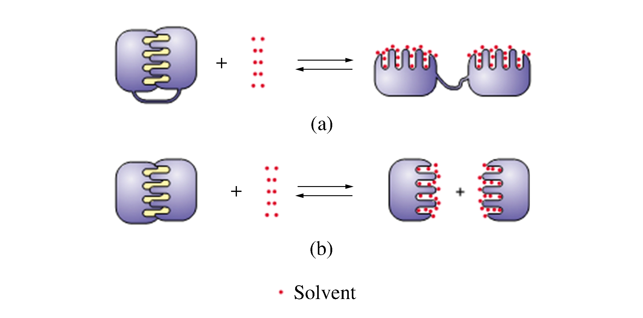
表 4 超高压对蛋白质四级结构的影响
Table 4. Influences of ultra-high pressure on the quaternary structure of proteins
Protein Pressure/(MPa) Changes of quaternary structure Test method Urate oxidase (Four
monomers)150-175 The molecule is dissociated reversibly
at 150 MPa and irreversibly at
175 MPa followed by aggregation[37]Fluorescence,
High pressure
crystallographyLactate dehydrogenases
(Four monomers)100-200 The molecule is dissociated into
monomers at 200 MPa[53]Fluorescence β dimer tryptophan synthase 80-240 50% of the molecules are dissociated
at 220 MPa, and the conformation is
recovered in 2-3 min after compression[54]Fluorescence Yeast jexokinase (Dimer) 0.1-240 The molecules are completely
dissociated at 200 MPa[55]Fluorescence GorEL (Tetradecameric) and
GorES (Heptameric) from
Escherichia coli50-300 GorES and GorEL dissociate at 250 MPa;
GorES reassociate readily after high
pressure release, but GorEL does not[56]Electrophoresis,
UV spectrumEnolase (A tripolymer with
three subunits α, β, γ)0.1-300 Molecules are dissociated into
two parts at 300 MPa[57]Fluorescence Lactose repressor protein
(Tetramer)0.1-300 Molecules begin to dissociate at 160 MPa
and completely collapse at 280 MPa[58]Fluorescence, Visible
spectrumTriosephosphate isomerase 0.1-350 50% of the proteins dissociate at 260 MPa
and completely collapse at 350 MPa[59]Fluorescence, Size-
exclusion FPLCYeast glyceraldehyde phosphate
dehydrogenase (Tetramer)0.1-280 Molecules begin to dissociate at 120 MPa
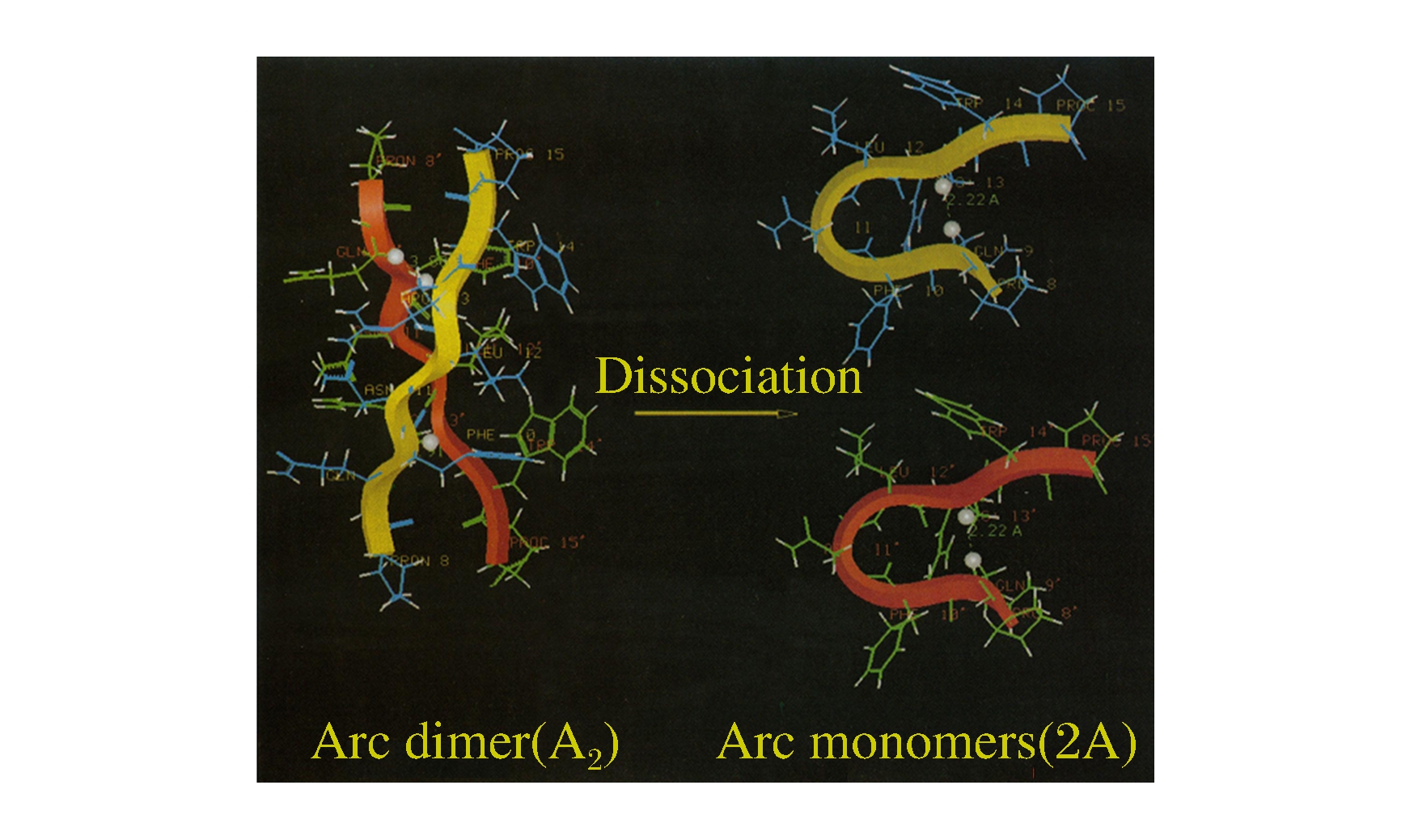
and completely collapse at 200 MPa[60]Fluorescence Arc repressor (Dimer) 0-500 Arc repressor begins to dissociate at 100-200 MPa
and completely collapses at 300-500 MPa[52, 61]Two-dimensional NMR Note:FPLC means fast protein liquid chromatography.
下载: 导出CSV
-
[1] 周林燕, 廖红梅, 张文佳, 等.食品高压技术研究进展和应用现状[J].中国食品学报, 2009, 9(4): 165-169. http://www.cnki.com.cn/Article/CJFDTotal-ZGSP200905029.htmZhou L Y, Liao H M, Zhang W J, et al. Review of high pressure technologies for food processing[J]. Journal of Chinese Institute of Food Science and Technology, 2009, 9(4): 165-169. (in Chinese) http://www.cnki.com.cn/Article/CJFDTotal-ZGSP200905029.htm [2] 廖小军.超高压技术在果蔬加工中大有可为[J].农业工程技术, 2009(9): 36-38. http://www.cnki.com.cn/Article/CJFDTotal-NYGN200909015.htmLiao X J. HHP has bright prospect in fruit and vegetable processing[J]. Agriculture Engineering Technology, 2009(9): 36-38. (in Chinese) http://www.cnki.com.cn/Article/CJFDTotal-NYGN200909015.htm [3] 上官丽娟, 马永昆, 崔凤杰, 等.高压处理对辣根过氧化物酶活性及构象的影响[J].高压物理学报, 2011, 25(5): 475-480. http://www.cqvip.com/QK/96553X/20115/39721767.htmlShangguan L J, Ma Y K, Cui F J, et al. Effects of high pressure processing on the activity and the conformation of horseradish peroxidase[J]. Chinese Journal of High Pressure Physics, 2011, 25(5): 475-480. (in Chinese) http://www.cqvip.com/QK/96553X/20115/39721767.html [4] 马汉军, 周光宏, 余小领, 等.高压与加热协同处理对牛肌肉中蛋白酶活性的影响[J].高压物理学报, 2011, 25(1): 89-96. http://www.cqvip.com/QK/96553X/201101/37267496.htmlMa H J, Zhou G H, Yu X L, et al. Effects of combined high pressure and thermal treatment on protease activities in beef muscle[J]. Chinese Journal of High Pressure Physics, 2011, 25(1): 89-96. (in Chinese) http://www.cqvip.com/QK/96553X/201101/37267496.html [5] 陈小强, 章银军, 张士康, 等.超高压处理对毛栓菌多酚氧化酶的影响[J].高压物理学报, 2012, 26(2): 235-240. http://www.cqvip.com/QK/96553X/201202/41757138.htmlChen X Q, Zhang Y J, Zhang S K, et al. Effect of high pressure processing on polyphenol oxidase from trametes trogii[J]. Chinese Journal of High Pressure Physics, 2012, 26(2): 235-240. (in Chinese) http://www.cqvip.com/QK/96553X/201202/41757138.html [6] Mozhaev V V, Heremans K, Frank J, et al. High pressure effects on protein structure and function[J]. Proteins Struct Funct Bioinf, 1996, 24(1): 81-91. doi: 10.1002/(SICI)1097-0134(199601)24:1<81::AID-PROT6>3.0.CO;2-R [7] Silva J L, Foguel D, Royer C A. Pressure provides new insights into protein folding, dynamics and structure[J]. Trends Biochem Sci, 2001, 26(10): 612-618. doi: 10.1016/S0968-0004(01)01949-1 [8] Boonyaratanakornkit B B, Park C B, Clark D S. Pressure effects on intra-and intermolecular interactions within proteins[J]. Biochim Biophys Acta, 2002, 1595(1/2): 235-249. http://europepmc.org/abstract/MED/11983399 [9] Eisenmenger M J, Reyes-De-Corcuera J I. High pressure enhancement of enzymes: A review[J]. Enzyme Microb Technol, 2009, 45(5): 331-347. doi: 10.1016/j.enzmictec.2009.08.001 [10] Gross M, Jaenicke R. Proteins under pressure: The influence of high hydrostatic pressure on structure, function and assembly of proteins and protein complexes[J]. Eur J Biochem, 1994, 221(2): 617-630. doi: 10.1111/j.1432-1033.1994.tb18774.x [11] 卜平宇, 夏泉.普通化学[M].北京: 科学出版社, 2009: 253.Bu P Y, Xia Q. General Chemistry[M]. Beijing: Science Press, 2009: 253. (in Chinese) [12] Bridgman P W. The Physics of High Pressure[M]. London: George Bell & Sons Ltd, 1931: 450. [13] Gekko K, Hasegawa Y. Compressibility-structure relationship of globular proteins[J]. Biochemistry, 1986, 25(21): 6563-6571. doi: 10.1021/bi00369a034 [14] Prehoda K E, Mooberry E S, Markley J L. Pressure denaturation of proteins: Evaluation of compressibility effects[J]. Biochemistry, 1998, 37(17): 5785-5790. doi: 10.1021/bi980384u [15] Vidugiris G J A, Royer C A. Determination of the volume changes for pressure-induced transitions of apomyoglobin between the native, molten globule, and unfolded states[J]. Biophys J, 1998, 75(1): 463-470. doi: 10.1016/S0006-3495(98)77534-4 [16] Seemann H, Winter R, Royer C A. Volume, expansivity and isothermal compressibility changes associated with temperature and pressure unfolding of Staphylococcal nuclease[J]. J Mol Biol, 2001, 307(4): 1091-1102. doi: 10.1006/jmbi.2001.4517 [17] Roche J, Caro J A, Norberto D R, et al. Cavities determine the pressure unfolding of proteins[J]. Proc Natl Acad Sci, 2012, 109(18): 6945-6950. doi: 10.1073/pnas.1200915109 [18] Visser A, Li T M, Drickamer H G, et al. Effect of pressure upon the fluorescence of various flavodoxins[J]. Biochemistry, 1977, 16(22): 4879-4882. doi: 10.1021/bi00641a020 [19] Zipp A, Kauzmann W. Pressure denaturation of metmyoglobin[J]. Biochemistry, 1973, 12(21): 4217-4228. doi: 10.1021/bi00745a028 [20] Kornblatt J A, Hui Bon Hoa G, Heremans K. Pressure-induced effects on cytochrome oxidase: The aerobic steady state[J]. Biochemistry, 1988, 27(14): 5122-5128. doi: 10.1021/bi00414a026 [21] Fuentes E J, Wand A J. Local stability and dynamics of apocytochrome b562 examined by the dependence of hydrogen exchange on hydrostatic pressure[J]. Biochemistry, 1998, 37(28): 9877-9883. doi: 10.1021/bi980894o [22] Collins M D, Quillin M L, Hummer G, et al. Structural rigidity of a large cavity-containing protein revealed by high-pressure crystallography[J]. J Mol Biol, 2007, 367(3): 752-763. doi: 10.1016/j.jmb.2006.12.021 [23] Abe F, Kato C, Horikoshi K. Pressure-regulated metabolism in microorganisms[J]. Trends Microbiol, 1999, 7(11): 447-453. doi: 10.1016/S0966-842X(99)01608-X [24] Heremans L, Heremans K. Raman spectroscopic study of the changes in secondary structure of chymotrypsin: Effect of pH and pressure on the salt bridge[J]. Biochim Biophys Acta, 1989, 999(2): 192-197. doi: 10.1016/0167-4838(89)90217-3 [25] Hei D J, Clark D S. Pressure stabilization of proteins from extreme thermophiles[J]. Appl Environ Microbiol, 1994, 60(3): 932-939. doi: 10.1128/AEM.60.3.932-939.1994 [26] Day R, García A E. Water penetration in the low and high pressure native states of ubiquitin[J]. Proteins Struct Funct Bioinf, 2008, 70(4): 1175-1184. doi: 10.1002/prot.21562 [27] Dadarlat V M, Post C B. Decomposition of protein experimental compressibility into intrinsic and hydration shell contributions[J]. Biophys J, 2006, 91(12): 4544-4554. doi: 10.1529/biophysj.106.087726 [28] 王镜岩.生物化学[M].北京: 高等教育出版社, 2002: 626.Wang J Y. Biochemistry[M]. Beijing: Higher Education Press, 2002: 626. (in Chinese) [29] Hayert M, Perrier-Cornet J M, Gervais P. A simple method for measuring the pH of acid solutions under high pressure[J]. J Phys Chem A, 1999, 103(12): 1785-1789. doi: 10.1021/jp983204z [30] Peng X, Jonas J, Silva J L. Molten-globule conformation of Arc repressor monomers determined by high-pressure 1H NMR spectroscopy[J]. Proc Natl Acad Sci, 1993, 90(5): 1776-1780. doi: 10.1073/pnas.90.5.1776 [31] Imai T, Sugita Y. Dynamic correlation between pressure-induced protein structural transition and water penetration[J]. J Phys Chem B, 2010, 114(6): 2281-2286. doi: 10.1021/jp909701j [32] Collins M D, Hummer G, Quillin M L, et al. Cooperative water filling of a nonpolar protein cavity observed by high-pressure crystallography and simulation[J]. Proc Natl Acad Sci, 2005, 102(46): 16668-16671. doi: 10.1073/pnas.0508224102 [33] Hédoux A, Guinet Y, Paccou L. Analysis of the mechanism of lysozyme pressure denaturation from Raman spectroscopy investigations, and comparison with thermal denaturation[J]. J Phys Chem B, 2011, 115(20): 6740-6748. doi: 10.1021/jp2014836 [34] Grigera J R, McCarthy A N. The behavior of the hydrophobic effect under pressure and protein denaturation[J]. Biophys J, 2010, 98(8): 1626-1631. doi: 10.1016/j.bpj.2009.12.4298 [35] Ando N, Barstow B, Baase W A, et al. Structural and thermodynamic characterization of T4 lysozyme mutants and the contribution of internal cavities to pressure denaturation[J]. Biochemistry, 2008, 47(42): 11097-11109. doi: 10.1021/bi801287m [36] Akasaka K, Li H, Yamada H, et al. Pressure response of protein backbone structure: Pressure-induced amide 15N chemical shifts in BPTI[J]. Protein Sci, 1999, 8(10): 1946-1953. doi: 10.1110/ps.8.10.1946 [37] Girard E, Marchal S, Perez J, et al. Structure-function perturbation and dissociation of tetrameric urate oxidase by high hydrostatic pressure[J]. Biophys J, 2010, 98(10): 2365-2373. doi: 10.1016/j.bpj.2010.01.058 [38] Le Tilly V, Sire O, Alpert B, et al. An infrared study of 2H-bond variation in myoglobin revealed by high pressure[J]. Eur J Biochem, 1992, 205(3): 1061-1065. doi: 10.1111/j.1432-1033.1992.tb16874.x [39] Kangur L, Timpmann K, Freiberg A. Stability of integral membrane proteins under high hydrostatic pressure: The LH2 and LH3 antenna pigment-protein complexes from photosynthetic bacteria[J]. J Phys Chem B, 2008, 112(26): 7948-7955. doi: 10.1021/jp801943w [40] Hummer G, Garde S, García A E, et al. The pressure dependence of hydrophobic interactions is consistent with the observed pressure denaturation of proteins[J]. Proc Natl Acad Sci, 1998, 95(4): 1552-1555. doi: 10.1073/pnas.95.4.1552 [41] Hemley R J. Effects of high pressure on molecules[J]. Annu Rev Phys Chem, 2000, 51: 763-800. doi: 10.1146/annurev.physchem.51.1.763 [42] Chen W, Heymann G, Kursula P, et al. Effects of gigapascal level pressure on protein structure and function[J]. J Phys Chem B, 2012, 116(3): 1100-1110. doi: 10.1021/jp207864c [43] Subirade M, Loupil F, Allain A, et al. Effect of dynamic high pressure on the secondary structure of β-lactoglobulin and on its conformational properties as determined by Fourier transform infrared spectroscopy[J]. Int Dairy J, 1998, 8(2): 135-140. doi: 10.1016/S0958-6946(98)00034-X [44] Ngarize S, Herman H, Adams A, et al. Comparison of changes in the secondary structure of unheated, heated, and high-pressure-treated β-lactoglobulin and ovalbumin proteins using fourier transform raman spectroscopy and self-deconvolution[J]. J Agric Food Chem, 2004, 52(21): 6470-6477. doi: 10.1021/jf030649y [45] Rouget J B, Schroer M A, Jeworrek C, et al. Unique features of the folding landscape of a repeat protein revealed by pressure perturbation[J]. Biophys J, 2010, 98(11): 2712-2721. doi: 10.1016/j.bpj.2010.02.044 [46] Takeda N, Kato M, Taniguchi Y. Pressure-and thermally-induced reversible changes in the secondary structure of ribonuclease: A studied by FT-IR spectroscopy[J]. Biochemistry, 1995, 34(17): 5980-5987. doi: 10.1021/bi00017a027 [47] 阎隆飞, 孙之荣.蛋白质分子结构[M].北京: 清华大学出版社, 1999: 334.Yan L F, Sun Z R. Structure of Proteins[M]. Beijing: Tsinghua University Press, 1999: 334. (in Chinese) [48] Knorr D, Heinz V, Buckow R. High pressure application for food biopolymers[J]. Biochim Biophys Acta, 2006, 1764(3): 619-631. doi: 10.1016/j.bbapap.2006.01.017 [49] Tschirret-Guth R A, Hoa G H B, de Montellano P R O. Pressure-induced deformation of the cytochrome P450cam active site[J]. J Am Chem Soc, 1998, 120(15): 3590-3596. doi: 10.1021/ja973909z [50] Tschirret-Guth R A, Koo L S, Hoa G H, et al. Reversible pressure deformation of a thermophilic cytochrome P450 enzyme(CYP119)and its active-site mutants[J]. J Am Chem Soc, 2001, 123(15): 3412-3417. doi: 10.1021/ja003947+ [51] Li H, Yamada H, Akasaka K. Effect of pressure on the tertiary structure and dynamics of folded basic pancreatic trypsin inhibitor[J]. Biophys J, 1999, 77(5): 2801-2812. doi: 10.1016/S0006-3495(99)77112-2 [52] Peng X, Jonas J, Silva J L. Molten-globule conformation of Arc repressor monomers determined by high-pressure 1H NMR spectroscopy[J]. Proc Natl Acad Sci, 1993, 90(5): 1776-1780. doi: 10.1073/pnas.90.5.1776 [53] King L, Weber G. Conformational drift of dissociated lactate dehydrogenases[J]. Biochemistry, 1986, 25(12): 3632-3637. doi: 10.1021/bi00360a023 [54] Silva J L, Miles E W, Weber G. Pressure dissociation and conformational drift of the beta dimer of tryptophan synthase[J]. Biochemistry, 1986, 25(19): 5780-5786. doi: 10.1021/bi00367a065 [55] Ruan K, Weber G. Dissociation of yeast hexokinase by hydrostatic pressure[J]. Biochemistry, 1988, 27(9): 3295-3301. doi: 10.1021/bi00409a026 [56] Panda M, Ybarra J, Horowitz P M. High hydrostatic pressure can probe the effects of functionally related ligands on the quaternary structures of the chaperonins GroEL and GroES[J]. J Biol Chem, 2001, 276(9): 6253-6259. doi: 10.1074/jbc.M009530200 [57] Paladini A A Jr, Weber G. Pressure-induced reversible dissociation of enolase[J]. Biochemistry, 1981, 20(9): 2587-2593. doi: 10.1021/bi00512a034 [58] Royer C A, Weber G, Daly T J, et al. Dissociation of the lactose repressor protein tetramer using high hydrostatic pressure[J]. Biochemistry, 1986, 25(25): 8308-8315. doi: 10.1021/bi00373a027 [59] Rietveld A W, Ferreira S T. Deterministic pressure dissociation and unfolding of triose phosphate isomerase: Persistent heterogeneity of a protein dimer[J]. Biochemistry, 1996, 35(24): 7743-7751. doi: 10.1021/bi952118b [60] Ruan K, Weber G. Hysteresis and conformational drift of pressure-dissociated glyceraldehydephosphate dehydrogenase[J]. Biochemistry, 1989, 28(5): 2144-2153. doi: 10.1021/bi00431a028 [61] Peng X, Jonas J, Silva J L. High-pressure NMR study of the dissociation of Arc repressor[J]. Biochemistry, 1994, 33(27): 8323-8329. doi: 10.1021/bi00193a020 -

 下载:
下载:

 点击查看大图

计量
- 文章访问数: 7231
- HTML全文浏览量: 2606
- PDF下载量: 297
