| Citation: | KANG Xu, LIU Jin. Phase Retrieval and Reconstruction of Coherent Diffraction Imaging[J]. Chinese Journal of High Pressure Physics, 2019, 33(3): 030105. doi: 10.11858/gywlxb.20190761

|

| [1] |
CHAO W, HARTENECK B D, LIDDLE J A, et al. Soft X-ray microscopy at a spatial resolution better than 15 nm [J]. Nature, 2005, 435(7046): 1210. doi: 10.1038/nature03719
|
| [2] |
BARBER J L, BARNES C W, SANDBERG R L, et al. Diffractive imaging at large fresnel number: challenge of dynamic mesoscale imaging with hard X-rays [J]. Physical Review B, 2014, 89(18): 184105. doi: 10.1103/PhysRevB.89.184105
|
| [3] |
XIAO X H, SHEN Q. Wave propagation and phase retrieval in fresnel diffraction by a distorted-object approach [J]. Physical Review B, 2005, 72(3): 033103. doi: 10.1103/PhysRevB.72.033103
|
| [4] |
MIAO J W, AMONETTE J E, NISHINO Y, et al. Direct determination of the absolute electron density of nanostructured and disordered materials at sub-10-nm resolution [J]. Physical Review B, 200, 68(1): 012201.
|
| [5] |
SAYER D. Some implications of a theorem due to shannon [J]. Acta Crystallographica, 1952, 5: 843.
|
| [6] |
GERCHBERG R W, SAXTON W O. A practical algorithm for the determination of phase from image and diffraction plane pictures [J]. Optik, 1972, 35: 237.
|
| [7] |
FIENUP J R. Phase retrieval algorithm: a comparison [J]. Applied Optics, 1982, 21: 2758. doi: 10.1364/AO.21.002758
|
| [8] |
ELSER V. Phase retrieval by iterated projections [J]. Journal of the Optical Society of America, 2003, 20(1): 40. doi: 10.1364/JOSAA.20.000040
|
| [9] |
CHEN C C, MIAO J, WANG C W, et al. Application of optimization technique to noncrystalline X-ray diffraction microscopy: guided hybrid input-output method [J]. Physical Review B, 2007, 76(6): 064113. doi: 10.1103/PhysRevB.76.064113
|
| [10] |
LUKE D R. Relaxed averaged alternating reflections for diffraction imaging [J]. Inverse Problems, 2005, 21: 37. doi: 10.1088/0266-5611/21/1/004
|
| [11] |
MARCHESINI S, HE H, CHAPMAN H N, et al. X-ray image reconstruction from a diffraction pattern alone [J]. Physical Review B, 2003, 68: 140101. doi: 10.1103/PhysRevB.68.140101
|
| [12] |
MIAO J, SYAER D, CHAPMAN H N. Phase retrieval from the magnitude of the fourier transforms of nonperiodic objects [J]. Josa A, 1998, 15: 1662. doi: 10.1364/JOSAA.15.001662
|
| [13] |
周光照, 佟亚军, 陈灿, 等. 相干X射线衍射成像的数字模拟研究 [J]. 物理学报, 2011, 60(2): 028701.
ZHOU G Z, TONG Y J, CHEN C, et al. Digital simulation for coherent X-ray diffractive imaging [J]. Acta Physica Sinica, 2011, 60(2): 028701.
|
| [14] |
VARTANYANTS I A, ROBINSON I K. Partial coherence effects on the imaging of small crystals using coherent X-ray diffraction [J]. Journal of Physics: Condensed Matter, 2001, 13(47): 10593. doi: 10.1088/0953-8984/13/47/305
|
| [15] |
MIAO J W, CHARALAMBOUS P, KIRZ J, et al. Extending the methodology of X-ray crystallography to allow imaging of micrometer-sized non-crystalline specimens [J]. Nature, 1999, 400: 342. doi: 10.1038/22498
|
| [16] |
MIAO J W, NISHINO Y, KOHNURA Y, et al. Quantitative image reconstruction of GaN quantum dots from oversampled diffraction intensities alone [J]. Physical Review Letters, 2005, 95(8): 085503. doi: 10.1103/PhysRevLett.95.085503
|
| [17] |
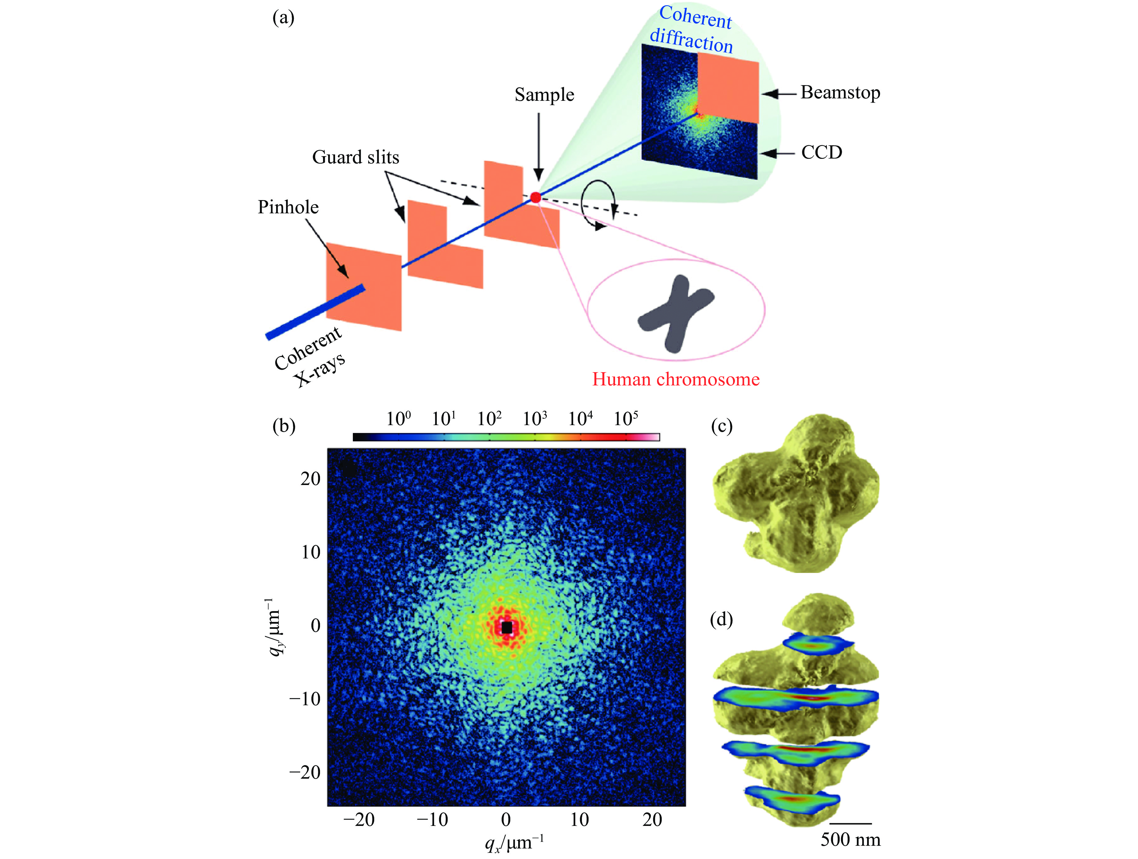
NISHINO Y, TAKAHASHI Y, IMAMOTO N, et al. Three-dimensional visualization of a human chromosome using coherent X-ray diffraction [J]. Physical Review Letters, 2009, 102(1): 018101. doi: 10.1103/PhysRevLett.102.018101
|
| [18] |
EKEBERG T, SVENDA M, ABERGEL C, et al. Three-dimensional reconstruction of the giant mimivirus particle with an X-ray free-electron laser [J]. Physical Review Letters, 2015, 114(9): 098102. doi: 10.1103/PhysRevLett.114.098102
|
| [19] |
DUANE N T, ELSER V. Reconstruction algorithm for single-particle diffraction imaging experiments [J]. Physical Review E, 2009, 80(2): 026705. doi: 10.1103/PhysRevE.80.026705
|
| [20] |
MIAO J W, CHEN C C, SONG C, et al. Three-dimensional GaN-Ga2O3 core shell structure revealed by X-ray diffraction microscopy [J]. Physical Review Letters, 2006, 97(21): 215503. doi: 10.1103/PhysRevLett.97.215503
|
| [21] |
TAKAHASHI Y, NISHINO Y, TSUTSUMI R, et al. High-resolution projection image reconstruction of thick objects by hard X-ray diffraction microscopy [J]. Physical Review B, 2010, 82(21): 214102. doi: 10.1103/PhysRevB.82.214102
|
| [22] |
THIBAULT P, DIEROLF M, MENZEL A, et al. High-resolution scanning X-ray diffraction microscopy [J]. Science, 2008, 321(5887): 379. doi: 10.1126/science.1158573
|
| [23] |
RODENBURG J M, HURST A C, CULLIS A G, et al. Hard-X-ray lensless imaging of extended objects [J]. Physical Review Letters, 2007, 98(3): 034801. doi: 10.1103/PhysRevLett.98.034801
|
| [24] |
KLAUS G, PIERRE T, SEBASTIAN K, et al. Quantitative biological imaging by ptychographic X-ray diffraction microscopy [J]. Proceedings of the National Academy of Sciences of the United States of America, 2010, 107(2): 529. doi: 10.1073/pnas.0905846107
|
| [25] |
DIEROLF M, MENZEL A, THIBAULT P, et al. Ptychographic X-ray computed tomography at the nanoscale [J]. Nature, 2010, 467(7314): 436. doi: 10.1038/nature09419
|
| [26] |
ROBINSON I K, VARTANYANTS I A, WILLIAMS G J, et al. Reconstruction of the shapes of gold nanocrystals using coherent X-ray diffraction [J]. Physical Review Letters, 2001, 87(19): 195505. doi: 10.1103/PhysRevLett.87.195505
|
| [27] |
WILLIAMS G J, PFEIFER M A, VARTANYANTS I A, et al. Three-dimensional imaging of microstructure in Au nanocrystals [J]. Physical Review Letters, 2003, 90(17): 175501. doi: 10.1103/PhysRevLett.90.175501
|
| [28] |
PFEIFER M A, WILLIAMS G J, VARTANYANTS I A, et al. Three-dimensional mapping of a deformation field inside a nanocrystal [J]. Nature, 2006, 442(7098): 63. doi: 10.1038/nature04867
|
| [29] |
NEWTON M C, LEAKE S J, HARDER R, et al. Three-dimensional imaging of strain in a single ZnO nanorod [J]. Nature Materials, 2010, 9(2): 279.
|
| [30] |
HARDER R, ROBINSON I. Coherent X-ray diffraction imaging of strain at the nanoscale [J]. Nature Materials, 2009, 8(4): 291. doi: 10.1038/nmat2400
|
| [31] |
GANG X, OUSSAMA M, MANFRED R, et al. Coherent X-ray diffraction imaging and characterization of strain in silicon-on-insulator nanostructures [J]. Advanced Materials, 2014, 26(46): 7747. doi: 10.1002/adma.v26.46
|
Figures(10)
Supported by: Beijing Renhe Information Technology Co. Ltd support:
info@rhhz.net


 DownLoad: